Mark Reed, DPM
DABFAS FAPWCA
714-528-3668


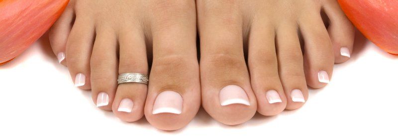

Bunion Deformity
Bunions Deformity Treatment!
Podiatrist Dr. Mark Reed has performed more than a thousand bunion corrections over 30+ years in being in practice. The Foot Doctor specializes in bunion corrective surgery with the emphasis of creating beautiful feet while minimizing the pain associated with these procedures. As a patient considering bunion surgery, you will not find a more compassionate, personal, credentialed, or experienced Podiatrist in the area.
In selecting a Podiatrist to perform your bunion surgery, experience should be a major consideration. Dr. Mark Reed emphasizes a conservative approach to bunion treatment by assessing the severity of the bunion on an annual basis with X-ray evaluation and working on trying to slow the progression of the bunion deformity using toe spacers, custom molded foot orthotics and motion control running shoes to name a few interventions. Bunion surgery is scheduled when there is pain, arthritic degeneration formation in the joint or the patient has a strong preference to correct the unsightly deformity based on what would be optimal for the patient's work and family priorities. There are many patients who have bunions where the Podiatrist have not seen their bunions increase in many years with conservative intervention. Other patients, based on their annual X-rays are not so fortunate and these patients are encouraged to consider surgery based on a six month to one-year window to find the most optimal time to least impact their schedules. Rarely is there ever a reason to rush into bunion surgery considering most patients have lived with the condition for many years.

Bunion Deformity - A Complex Deformity
The history of bunions has been discussed in the medical podiatry literature for more than 100 years. The term "bunion" is derived from the Latin word for turnip. Bunions usually occur on both feet with one foot usually worse than the other. In western countries, bunions occur more in women due to the type of shoes women wear. In countries where men and women do not wear shoes, the incidence of bunions in men and women has been found to be the same. However, the person's inherited or developed tendency to walk flat-footed has been shown to be the primary cause of bunion deformities. In rare circumstances, neurological disorders, rheumatoid arthritis, and developmental deformities can also cause a bunion deformity.
The classic bunion is medically known as Hallux Abductovalgus or HAV. It is described as a bump on the side of the great toe joint. This bump represents an actual deviation of the First metatarsal bone and often an overgrowth of bone on the metatarsal head. In addition, there is also deviation of the great toe toward the second toe. In severe cases, the great toe can either lie above or below the second toe. Shoes are often blamed for creating these problems. This, however, is inaccurate. It has been noted that primitive tribes were going barefoot is the norm will also develop bunions. Bunions develop from abnormal foot structure and mechanics (e.g. excessive pronation), which place an undue load on the 1st metatarsal. This leads to stretching of supporting soft tissue structures such as joint capsules and ligaments with the end result being gradual deviation of the 1st metatarsal. As the deformity increases, there is an abnormal pull of certain tendons, which leads to the drifting of the great toe toward the 2nd toe while the first knuckle joint (metatarsophalangeal joint) drifts away from the foot and become prominent on the side of the foot. As the bunion increases, there is also adaptation of the joint itself that occurs due to internal pressures that injures the cartilage and progressive degenerative joint disease occurs commonly known as arthritis.
One of the more common conditions treated by podiatric surgeons is the painful bunion. Patients with this condition will usually complain of pain when wearing certain shoes, especially snug fitting dress shoes, or with physical activity, such as walking or running. Bunions are most commonly treated by conservative treatments. This may involve shoe gear modification, more stable shoes, shoe padding, toe spaces and / or foot orthoses. When conservative interventions fail to provide adequate relief, surgery is often recommended. There are several surgical procedures to correct bunions. Selection of the most appropriate procedure for each patient requires knowledge of the level of deformity, review of the x-rays and an open discussion of the goals of the surgical procedure. Almost all surgical procedures require repositioning the first metatarsal head next to the second metatarsal head. In the case of mild to moderate bunion deformity, the bone correction is performed at the neck of the metatarsal (near the joint) to move the joint back next to the second joint or metatarsal head.
Symptoms Related to Bunion Deformity
The most common symptoms associated with this condition are pain on the side of the foot. Shoes will typically aggravate bunions. Stiff leather shoes or shoes with a tapered toe box is the prime offenders. This is why bunion pain is most common in women whose shoes have a pointed toe box. The bunion site will often be slightly swollen and red from the constant rubbing and irritation of a shoe. Occasionally, corns can develop between the 1st and 2nd toe from the pressure the toes rubbing against each other. On rare occasions, the joint itself can be acutely inflamed from the development of a sack of fluid over the bunion called a bursa. This is designed to protect and cushion the bone. However, it can become acutely inflamed, a condition referred to as bursitis.
Evaluation of a bunion deformity begins with obtaining a complete history and physical as well as obtaining X-rays of both feet. Typically, as a bunion deformity progresses, calluses form under the ball of the foot, a reddened area appears over the prominent first metatarsal head, lesser toe deformities appear, shoes no longer fit properly, degenerative joint disease or osteoarthritis changes occur, and the cosmetic appearance of the foot deteriorates.

BUNION SURGERY CONSIDERATIONS
Treatment of a bunion deformity primarily involves two issues, correcting the underlying cause of the bunion deformity, and when required, surgically realigning the bunion deformity. In cases where the bunion deformity is mild to moderate and the patient is not in pain, custom molded foot orthotics and toe spaces are usually prescribed to help correct the weight bearing forces which usually cause the bunion deformity and align the tendons pulling on the great toe.
The surgical correction of bunions is dependent upon the severity of the deformity, the patient's over-all health and activity level. Age and conditions such as diabetes do not preclude bunion surgery as a form of treatment.
There are several different approaches to the surgical correction of bunions. Most commonly, the surgery is performed in the area of the big toe joint. The bony prominence is removed, and the bone is surgically fractured to allow realignment of the joint and straightening of the big toe joint. This surgery is designed so that the patient can walk on the foot almost immediately following the procedure; however, activity must be significantly curtailed for several weeks following the surgery. Typically, the patient is instructed to remain home from work for at least one week with the foot propped up and elevated above the heart throughout the day. If the patient's job requires much standing or walking, they may be required to stay home from work for as much as six weeks. Often the patient may return to work sooner if they are placed in a removable below-the-knee walking cast. There are no short cuts to the healing time. Healing time is based upon basic physiological principles that are common to all human beings. Certain vitamins and nutrients may help with the healing process. Laser surgery does not alter the healing time and provides no significant advantage to the performance of the surgery.
Surgical Correction of Severe Bunion Deformity
If the bunion is more sever in nature surgery is performed further back on the bone in order to straighten the big toe. When surgery is performed in this area of the bone, there is greater instability of the bone after it is cut and moved into a corrected position. Generally, the surgeon will require the patient to wear a below-the-knee cast and use crutches for three to eight weeks. Initial bone healing takes six to eight weeks. This period of time can take longer in people who smoke.
The overall success rate and satisfaction of patients who have had bunion surgery is quite high. The most common complaint of patients is the healing time. This is particularly true if the patient is not adequately prepared or informed as to what to expect. Most patients experience minimal pain following the procedure and this pain is easily controlled with pain medication prescribed by the surgeon.
Possible Complications
Potential complications associated with the bunion corrective surgery are infection, swelling, pain, big toe shortening, arthritis degenerative changes, over or under-correction of the bunion, joint stiffness, delays in healing or non-healing of the bone, or healing of the bone in the wrong position. Most of these complications can be avoided by following the surgeon's instructions. Walking on the foot without the protection of a post-operative shoe or cast, or against the Surgeon's advice can lead to a dislocation of the bone where it has been cut. This results in delays in healing, non-healing of the bone or healing of the bone in the wrong position. Allowing the bandage to get wet increases the risk of infection. The most critical time for an infection to occur is within the first three days following surgery. Infection can also occur following this period of time but is less common.
Joint stiffness following bunion surgery is common, but generally improves with time. Postoperative physical therapy is useful to improve the movement of the joint but is not always necessary.
Bunions on Both Feet- Considerations with Regard to Surgery
If a person has bunions on both feet, many surgeons feel that their patients recover quicker and with fewer complications if the surgery is performed on one foot at a time. Many surgeons prefer to wait a minimum of four to five weeks between surgeries. Other surgeons prefer that their patients wait longer between surgeries.
Place of Service and Anesthesia Considerations
Most often the bunion surgery is performed in an outpatient surgery center or hospital. Some surgeons will perform this procedure in their office. Anesthesia for the surgery can range from a straight local anesthesia, given by injection into the area of surgery, to a general anesthesia with the administration of an anesthetic gas. A very common form of anesthesia is a combination of a local anesthesia and medicine given intra-venous to make the patient drowsy. This is commonly called twilight anesthesia.
Generally, there is very little blood loss during surgery. Most often the surgeon will use some form of tourniquet to stop bleeding during surgery. Because the surgery can be performed in a relatively short period of time the use of a tourniquet is very safe. Technically, the tourniquet can be left in place for as long as 90 minutes safely in most cases. Surgeons who perform bunion surgery are very knowledgeable in the use of tourniquets. The potential for the need for a blood transfusion with bunion surgery is nearly non-existent.
Can My Bunion Come Back?
It is important to understand that bunion surgery does not correct the cause of the bunion. Therefore, there is the possibility that the bunion can reoccur. How quickly a reoccurrence will occur is difficult to predict. It may take several years or just a matter of months for the bunion to begin to come back. Bunions are caused by abnormal movement of a set of joints below the ankle joint in the foot called the subtalar joints. To help prevent the bunion from reoccurring the patient should be prescribed a functional orthotic. These are custom-made shoe inserts that correct the abnormal function of the foot. Generally, they will fit in normal shoes without requiring the use of larger shoes. Most foot surgeons will suggest the use of orthotics following bunion surgery to help prevent the reoccurrence of the deformity.
BUNION SURGERY OVERVIEW
Surgical realignment of a bunion deformity consists of a realignment of the great toe and first metatarsal. In some cases where arthritic joint degeneration has occurred, besides realigning the joint deformity, the joint must also be fused or artificially replaced. A bunion deformity actually involves two unique deformities. The first is the widening of the knuckle joint of the great toe from the knuckle joint of the second toe and is called Metatarsus Primus Adductus. Metatarsus Primus Adductus is measured by measuring the angle created by drawing a bisection of the first and second metatarsal bones on a weight-bearing AP X-ray. The angle of the first metatarsal to the second metatarsal is called the 1-2 Inter-metatarsal Angle and usually is normal if under 10 degrees. The second deformity is the rotation of the great toe towards the second toe that is called the Hallux Abductovalgus Deformity.
In evaluating a patient for surgical correction of their bunion deformity, ultimately, the different causes of the patient's Metatarsus Premus Adductus and Abductovalgus Deformity must be determined by the surgeon. Usually, the cause of the bunion deformity is solely due to widening of the first metatarsal away from other metatarsals. However, there are many anatomical reasons that a bunion deformity can be in existence, and this is why a foot specialist should evaluate a bunion deformity prior to surgery. In correcting a bunion deformity, some doctors still cut off a portion of the knuckle joint of the great toe to narrow the appearance of the foot but do not realign the knuckle joint of the great toe. This type of narrowing procedure typically severely narrows the knuckle joint of the great toe and has been associated with numerous complications such as early degenerative joint arthritis. Typically, these narrowing procedures are temporary in giving any relief to the patient and other surgical procedures must be performed subsequently to realign the knuckle joint of the great toe. A second opinion should be obtained from a foot specialist if a doctor recommends a narrowing procedure that intends to narrow the knuckle joint of the great toe without realignment of the joint.
In discussing recovery expectations, in the hands of a good foot surgeon, for the patient with a mild to moderate bunion deformity that involves only the widening of the first metatarsal compared to the second metatarsal, the patient will have usually a two-to-four-week recovery period before returning to shoes and can walk on the foot during recovery. For the patient with a severe bunion deformity that involves only the widening of the first metatarsal from the second metatarsal, surgery usually involves an eight-to-ten-week period in which no weight bearing can occur.
In making a pre-operative evaluation of the patient, weight bearing X-rays must be evaluated to determine he appropriate procedures that need to be performed. The amount of angle between the first and second metatarsal is one of the main factors in determining which type of procedure is indicated and how much recovery time will be experienced by the patient. The angle between the first and second metatarsals is important because there is a point that the first metatarsal head cannot be sided back gains the second metatarsal. When the first metatarsal is too wide to slide it over against the second metatarsal, a wedge procedure must be performed to swing the metatarsal over far enough to move the first metatarsal head against the second metatarsal head. In evaluating X-rays, there are actually 11 other X-ray measurements that are typically evaluated in deciding what is the correct procedure for correcting a bunion deformity.
Early medical intervention using custom molded functional foot orthotics, silicone toe spacers, and motion control running shoes can in many cases prevent bunion surgery or slow the progression of the deformity for a lifetime. The use of foot orthotics after bunion surgery is important in preventing the return of the bunion deformity because the surgery only realigns the joint back next to the second metatarsal and does not actually correct the underlying cause of the bunion in how your naturally walk. Please review the foot orthotic article for information on foot orthotics. The use of a silicon toe spacer reduces stress to the ligaments of the joint and in many cases can return the patient to a pain free joint unless there is significant arthritic degeneration in the joint.
The best approach in deciding what to do about a developing bunion is to get an X-ray early in the progression of the deformity to gain an understanding of what is the best intervention to either prevent the deformity from increasing in severity. When there is pain, arthritic degenerative changes or a deformity that is progressing in severity, surgery should be considered but only after conservative interventions have been utilized.
Bunion Surgery - Head Procedures
First metatarsal neck osteotomies are known by various names based on the individual who first described the procedure (e.g. Austin, Reverdin-Green, Kalish-Austin). Regardless of the procedure, the goal of all these procedures is the same, to remove the bump and realign the joint. The first part of all bunion procedures involves removing the bump of bone from the side of the 1st metatarsal head. This is performed in a manner so as not to damage the viable part of the joint and not to leave any irregularities of bone that can cause future irritation in shoes. Once this is completed, the podiatric surgeon will create an osteotomy (bone cut) through the first metatarsal that will allow shifting the bone and realigning the joint. Depending on the type of osteotomy, the actual shape of the bone cut can vary. In the case of the Austin bunionectomy, the bone cut is V-shaped with the "V" sitting on its side and the tip of the "V" pointing toward the joint. When this cut is completed, the head of the metatarsal and joint is shifted toward the 2nd toe. In this way the bone and joint are repositioned in a more normal position. The Reverdin-Green osteotomy is made in a similar location but is trapezoidal in shape rather than V-shaped. Both these procedures are stable bone cuts and provide good correction of mild to moderate deformities. The Kalish-Austin bunionectomy is a modification of the Austin bunionectomy. It also is a V-shaped bone cut but is typically used for greater degrees of bunion deformities.
Because bone is cut and repositioned, it is often preferred to fixate or hold the bone in place with some external device. In the case of the Austin and Reverdin-Green osteotomies, this is most often accomplished by the use of a stainless-steel pin across the bone cut. This prevents accidental displacement and loss of correction. Over the past 5 years, it has become increasing more advantageous to use small stainless steel or titanium screws to provide compression of the bone and to hold the bone in position. This is the main advantage of the Kalish-Austin bunionectomy. By using the screws, bone will heal faster and will allow for earlier ambulation. The screws are typically left in permanently unless they cause irritation of the soft tissues while the pins are generally removed in the office setting in three to four weeks following the day of surgery. The surgery is generally preformed as an outpatient in a hospital or outpatient surgery center. Anesthesia is the choice of the surgeon made in consultation with the patient and anesthesiologist. Anesthesia may be a general anesthesia, twilight anesthesia or a local anesthesia.
Post Operative Care
The postoperative course and rehabilitation following bunion surgery depends on the procedure and can vary amongst podiatric surgeons. Patients have varying levels of postoperative pain but quite often the pain is significantly less than what the patient anticipates. A period of total non-weight bearing with crutches may be recommended in the first 3 to 5 days. In many instances, the surgeon may allow the patient to bear full weight in a postoperative surgical shoe. In all cases patients are instructed to limit their activities and to elevate their feet above their heart during the first 3 to 5 days. After this, a resumption of gradual weight bearing with a special surgical shoe is begun. Walking without the postoperative shoe is strictly prohibited. In cases where a pin is used, return to full weight bearing with a stiff soled walking shoe is allowed after the pin has been removed, generally in 3 to 4 weeks following the bunion surgery. Screws provide increased stability when used to fixate bone cuts and most patients can return to full weight bearing and regular shoes in 3-4 weeks following the surgery. The postoperative and rehabilitative course is improved by the use of ice and elevation of the extremity as much as possible. One of the most important aspects of the postoperative treatment is early motion of the joint to prevent joint stiffness. In most cases, range of motion exercises are begun almost immediately following surgery. No matter what the form of bone fixation is used, pins or screws; bone healing will take 6 to 8 weeks or longer. During this period of time, it is important that the patient not walk without shoes or in thin-soled shoes or sandals. Should the patient risk walking without an adequately supportive shoe, they risk re-fracturing the bone and increase the duration of healing.
Possible Complications
Complications following bunion surgery are uncommon but may include infection, suture reaction, delayed or nonunion of the osteotomy, irritation from the pin or screws, stiff joint or recurrence of the deformity. Recurrence of the deformity can be halted or slowed with the use of functional foot orthotics. It is important to realize that surgery does not correct the cause of the bunion deformity. Functional foot orthotics however do address the cause of the deformity and their use are strongly encouraged following bunion surgery. A rare complication is the over correction of the bunion deformity. This condition, called Hallux Varus, may require additional surgery for its correction.
Bunion Surgery - Shaft Procedures
Hallux valgus or bunion deformities have may different surgical techniques for their correction. One group of procedures that your surgeon may use is the shaft osteotomies. These osteotomies are different from the head osteotomies and also the procedures performed at the base of the metatarsal or at the metatarso-cuneiform joint, because they are performed in the middle of the first metatarsal.
The shaft osteotomies were designed to use internal fixation (screws) and to correct larger deformities. In most of these cases, your surgeon will use 2 screws to fixate the osteotomy. The osteotomy is longer than the head procedures and has more inherent stability because of more bone contact. Also, these procedures can correct larger deformities then the head procedures and about the same deformities as the base procedures.
There are two basic shaft osteotomy procedures that your doctor may talk to about: The Z bunionectomy or the offset V bunionectomy. These osteotomies are very similar and are used interchangeably, based on different patient characteristics, by most surgeons that perform these procedures. The decision to use these procedures over other procedures is typically surgeon preference. In most cases, these procedures are used for patient with mild to severe structural bunions without hypermobility. In old patients with poor bone stock, the surgeon may opt for other procedures.
What is the post-operative course?
Typically, the patient is allowed to bear weight immediately after surgery in a surgical shoe. Some doctors may have you use crutches for one to two weeks or use a slipper cast. This is surgeon's preference. It is not unusual for the front part of your foot to look bruised after the surgery. So, at the first dressing change, do not be surprised if your toes and the top of your foot are bruised. This will dissipate in 3-6 weeks. At two weeks after surgery, the sutures are typically removed and at three weeks most patients are advanced into a surgical shoe. After the first or second week, your surgeon may have you start range of motion of your big toe joint. It is important that you follow your doctor's instructions on all range of motion exercises to help return motion to the operative foot. As with all surgery on the foot and ankle, the limiting factor to advance into different shoe gear is swelling. This swelling can last up from 6 months to one year after surgery. Typically, most patients returned to pre-operative dress shoes in 6 to 8 weeks after surgery.
With any surgery, complications are possible. Every procedure has unique complications, and your surgeon will discuss these with you before surgery. Make sure that you ask any questions that you have about the surgery with your surgeon.
Keller Bunionectomy
Pain or discomfort in the great toe joint is a common occurrence amongst people seeking podiatric treatment. There are numerous reasons why people may experience pain or discomfort in this region. Pain in this area may be due to a restriction of motion, a condition referred to as hallux limitus or rigidus. This condition can lead to jamming of the joint and potential degenerative joint disease or arthritis. Ironically, this will lead to even further stiffening of the joint and pain with walking. Bunions or hallux abductovalgus deformities can also cause pain in the great toe joint. After years of this abnormal alignment of the joint arthritic changes can occur causing even more pain. Prior injury to the joint can lead to the development of traumatic arthritis. This is another potential cause of pain in the great toe joint. Patients with diabetes may develop an altogether different problem related to this lack of motion. In the presence of peripheral neuropathy (lack of painful sensation), these patients can develop skin breakdown and ulceration.
As you can see there are numerous causes for a painful joint. There are also numerous options, conservative and surgical, to treat these conditions. A Keller Arthroplasty is a surgical procedure designed to eliminate pain and discomfort in this joint. It is typically reserved for cases of severe arthritis, previous failed surgeries, diabetic ulcerations or certain types of bunion deformities.
Indications For Surgery
As with all surgical procedures there are certain criteria that are followed in choosing one procedure over another for any individual patient. In the case of the Keller arthroplasty, it is most commonly reserved for patients over the age of 55 with limited athletic activity. These patients are best able to tolerate the alteration in toe function created by this procedure. There should also be a moderate to severe amount of pain on movement of the joint, either passively or with walking that is not relieved by shoe gear, orthotics or other non-surgical means. X-rays are helpful to evaluate the condition of the bone and joint. These may show joint space narrowing, bone spurs or joint deterioration. As with any surgery, it is important that the patient have a clear understanding of all available options. They should also be aware of what to expect after surgery.
The Surgical Procedure
The procedure itself is fairly straightforward. An incision is made over the great toe joint. Once the joint is exposed, a small portion of bone is removed from the base of the proximal phalanx. This allows for an increase in motion in the joint and a reduction in pain. The defect created by the removal of the bone will fill in with soft tissue, creating a “false joint”. Some surgeons may choose to place a pin across the joint to maintain the position of the toe and to allow for scarring. The pin is usually left in place for 3-4 weeks. The soft tissue structures are then re-attached, and the wound is closed. The patient is then placed in a surgical shoe. Casting is not necessary and limited ambulation is usually allowed following this procedure.
What to Expect after Surgery
The postoperative recuperation usually involves use of the surgical shoe for 2-3 weeks. Limited ambulation may be allowed. If a pin was inserted, this is usually removed after 3-4 weeks. Because the pin exits out the tip of the great toe, it can usually be removed in the office. It does not require a second surgical procedure. Once the pin is removed, the patient can get the foot wet, increase their weightbearing activities, begin range of motion exercises and gradually advance to sneakers. Most people can return to their usual shoe gear and activity at 6 weeks.
The most common postoperative concerns are prolonged swelling. It is not unusual for some degree of swelling to persist beyond 3 months. This will typically resolve on its own. Occasionally, the use of a compression sock will expedite resolution of the swelling. Also, an orthotic device may be helpful to allow for more efficient transfer of weight during ambulation and more even distribution of weightbearing forces. All in all, when the preoperative criteria are met, this procedure can provide a significant degree of relief from a painful great toe joint.
Bunion Glossary of Terms

Mark Reed, DPM
DAPFAS FAPWCA
1275 N. Rose Drive, Suite 136
Placentia, CA 92870
Open M-F 8:30 to 5
Saturday: By Appointment
Info@Podiatry.Care
Fax: 714-528-0739
Office: 714-528-3668
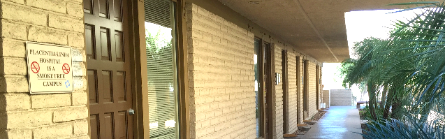
Located in the
Placentia Linda Hospital
Medical Building
Follow Dr. Mark Reed

*****
Beckie Burcham.
An absolutely amazing and genuine doctor who is not interested in only treating the symptoms, but rather treating the real cause! Dr. Reed explains everything in real simple terms that anyone can understand, especially seniors. He has amazing, common sense solutions (i.e. treatments) for unresolved diabetic foot ulcers where others have only wanted to continue cutting weekly into my family member's foot! Dr. Reed is absolutely AMAZING! And if you are getting no where with your currents foot issues or someone continues to only cut, please, please...do yourself a favor and contact Dr. Reed's office right away! Trust me, my family member has been dealing with an unresolved diabetic foot ulcer since 2018, and in just 3 weeks the foot is healing fantastic!!! Thank you Dr. Mark Reed. You have helped us to save the foot!!!
What an Amazing Podiatrist Dr. Mark Reed is! After years of other foot doctors just cutting into my family member's foot (diabetic foot ulcer), with numerous infections along the way...I did research and found Dr. Reed. Absolutely the best decision I have made for my loved one! After only 3 weeks of treatment, the foot ulcer is healing!!! No amputation! No continuous cutting and keeping the wound open and susceptible to infections! Brilliant man with great knowledge, kind heart, straight to the point and tells the truth the way it is! Dr. Reed does not just treat the symptoms, but rather the source of the problem! My family member's glucose levels are down to an average of 112 now. The wound is healing and new skin is growing!!! Amazing! I would highly recommend Dr. Reed to anyone and everyone! Thank you Dr. Reed, from the bottom of my heart! You have been a God send! Thank you!
*****
Mathew
H. I went to Dr. Reed because I had a chronic (5 months) wound on my leg that needed expert medical attention. Dr. Reed assessed the wound and knew how to treat it. He explained the process of healing the wound and what I needed to do in order to make the plan work.
I appreciated that Dr. Reed took the time to explain the process and his approach to me. As I followed the plan, I saw improvement within the first week and continued improvement in the following weeks. Within six weeks of my first appointment, the wound was healed.
Dr. Reed is personable and I enjoyed speaking with him in conversation. Most importantly, he told me what the treatment plan was, why it was that, and what I needed to do to ensure that it is effective. I am reluctant to go to doctors. I highly recommend Dr. Reed because he understands that many people feel that way and most importantly because the treatment was successful.
Lastly, the office staff are professional and kind. Dr. Reed's office is a first class group and I recommend them very highly.
*****
Paul Gordy. Dr. Reed is an outstanding podiatrist...he is state of the art medicine. This is by far the most kind, compassionate and caring physician who takes personal interest in his patients care and success in healing...when Care More and BlueShield doctors said they could not heal me...Dr. Read said "that's crazy", I'll heal you in 90 days .....and he DID!!!!!,
Amazing skill. You will not find a better person or doctor anywhere...
My family and I. Love Dr Reed.
You will too!
Paul Gordy
Yorba Linda Ca
*****
David M. The entire staff is friendly and very helpful. Dr. Reed is dedicated, spends time to take care of his patients. I had gangrene in my toe and Dr. Reed was diligent in his care and was able to eradicate the problem. I have recommended him to others, who had similar positive experiences.
***** Lee Godden. I have nasty bunions which eventually led to a painful stress fracture of the second metatarsal while playing basketball 2x/week. Dr. Reed managed my entire recovery, from initial visit to discharge two months later. He took X-rays and did ultrasound therapy. I was happy that his focus was on realignment and patient education, as opposed to surgery. I'm now running again thanks to the orthotic insoles, toe spacers, etc. that Dr. Reed put me in. I highly recommend Dr. Mark Reed.
***** Yvon Cariou. Dr. Reed did a great job on that ingrown toe nail, a painful condition. It took some time but Dr. was persistent and supported the process with detailed explanations and supplemental care as needed. A good office to go to.
***** Sue Alexander. If you’re looking for a podiatrist (foot and ankle doctor), may I suggest Dr. Mark Reed in Yorba Linda. Throughout my treatment Dr. Reed explained what he would be doing and why. Thank you Dr Reed for your expertise, kindness and encouragement! Sincerely, Sue Alexander
***** Salam Hamad. Hat off for Dr. Reed. He was the 4th doctor, second specialist to consult for my Plantar Fasciitis case. I went to him after the 1st specialist, using injections, boots and what not decided to operate on my heel in 30 days if the pain does not go away. Doctor Reed threw all that talk away and dismissed every recommendation. I went to him armed with lot of documents, dates and details of what the first 3 doctors did. He said, don't give the history, just tell me where it hurts. He examined the heel and the foot and diagnosed the problem immediately. He then prescribed the method to treat it. He did it! yes, it took more time than he thought but he DID it. I owe doctor Reed and appreciate him, his knowledge, experience and his caring attitude every time I walk. Yes, walking became possible for what he did. Oh! by the way, he is not in it for the money.
***** Sue Alexander. If you’re looking for a podiatrist (foot and ankle doctor), may I suggest Dr. Mark Reed in Yorba Linda. Throughout my treatment Dr. Reed explained what he would be doing and why. Thank you Dr. Reed for your expertise, kindness and encouragement! Sincerely, Sue Alexander
***** Monsie Crane.
Dr Reed cured me of the neuropathy in my toes. The neuropathy was a result of chemotherapy, which I received as a cancer treatment. He also helped me with proper shoe fitting for bunions and longer second toes. I highly recommend Dr Reed.
***** Colleen Livermore. Dr Mark Reed has been an excellent doctor, evaluating my foot and ankle pain, and giving me the instruction and exercises needed to strengthen and heal. I appreciate his excellence, expertise and experience. Ankle pain can be so challenging so to feel better after his help is a great relief.
Reply1
*****
Matthew Jones.
Dr. Reed has been my podiatrist for approximately 2 years now. He remains one of my favorite doctors because of how kind he is, how punctual he is and most importantly how effective he is.
I have been to several other podiatrists in the past and I just about gave up until I found Dr. Reed on google. The results he has given me have changed the way I feel and the way I look.
***** Lily Lambutu. Dr. Reed is an excellent doctor. He listens, gets straight to the point, explains everything, and pays attention to detail. He answers all questions honestly. I've seen him for ingrown toenails and a tumor on the bottom of my foot. I trust him completely as he takes the time to assess the issue and will take a conservative approach if it will solve the problem.
***** Bryant Brislin. Dr. Reed took a thoughtful approach to my issue and came up with a solution that other podiatrists had not. He took his time and was very friendly, which was refreshing. He seemed to truly care for me to get better!
***** Lisa Fogerty.
Dr Reed is an outstanding doc. Several years ago I had a hammer toe surgery done by another doctor who damaged the nerves in my foot. I was in pain for 3 years and was told by that doctor that I would have to live with it and that eventually the nerves would heal. They didn't. I couldn't sleep due to the pain and had to alter my exercise routine. Dr Reed took on the case, correctly diagnosed the problem and started a program of injections to get the nerves to regenerate. He told me to be patient and that "we" would fix the problem. He always kept me informed of the procedures and made me part of treatment plan. It took several months but today my foot and toes are healed thanks to Dr Reed's persistence.
My wife dislocated several toes in an accident and was in significant pain. Dr Reed fixed her up as well.
We are both very grateful to Dr Reed and his excellent staff for their kind and professional medical services and highly recommend him. If you want the best podiatrist in Orange County you found him in Dr Mark Reed.
***** Samantha Smith. It’s been almost 2 years since Dr. Reed cut off an amputation stump neuroma and then reconnected my husbands medial plantar nerve. This man has changed our lives forever. After being turned down and rejected by SO many doctors, we had no hope left...then we found Dr. Reed. He really did the impossible! My husband was in so much pain for so long and now feels little to no pain thanks to our friend Dr. Reed. We owe everything to him and will always be SO thankful for everything he has done for us. He will forever be a huge part of our lives.
Reply1
***** Leo Burke. I'm an avid hiker but have been plagued for years by a large toenail that was sensitive to pressure and would have to go to great lengths to avoid losing the nail. But every now and then I would lose the nail and all this was impacting my hiking (and ruining some vacations). I was seeing other podiatrists and we would try various work-arounds, but I finally decided that I would get rid of that nail once and for all. I had a lot of trepidation about that, but I decided to see Dr. Reed to consult with him on doing so. He assured me that it would be no problem at all, which surprised me, so I agreed to have him do the removal. Amazingly, it turned out to be a piece of cake with little to no pain, and I was back in action very quickly. It pains me to realize that if I had seen him a long time ago I could have avoided years of aggravation and frustration. Not only that, but he's an expert on hiking boots and given me a lot of help in boot selection.
He's been in this for so long that he's a fountain of knowledge and enjoys imparting his wisdom to his patients. For example, I also have a pressure sensitive small toe on my other foot and he was able to give me samples of some products that can relieve pressure on the toe that I hadn't been aware of. If that doesn't do it, I can cut an "X" in the boot over that area and cover it with a piece of leather. He then supplied me with a piece of leather that could be used for that purpose and told me what glue to use.
There's no question about it - Dr. Reed is the man to see for your foot problems. He's head and shoulders above everyone else.
***** Gloria Gonzalez. Dr. Reed is a great podiatrist. He's the only doctor that has helped my family's and my foot issues. He didn't suggest surgery for my issue, and his solution was easy and innovative. A couple of years later, and his solution still works.
Reply1
***** Nikki Clark. I have been a patient of Dr. Reed's for 6 months. During this time I have found both his professional staff and Dr. Reed to apply the utmost patience, compassion and expertise. They always make time for appointments and during doctor visits. Dr. Reed has an impeccable bedside manner, provides all options available and genuinely cares for a positive solution. Dr. Reed is a student of his practice and applies current medical advancements. I would recommend Dr. Reed and his staff to family and friends for problems they might be experiencing.
*****
Viet Tieu Bao: I have a great experience with Dr. Reed. First of all, he's an awesome surgeon as it turned out i didn't have any pain after my surgery (i had a tumor that grew next to my Achilles tendon that needed to be removed and in order to have a clean removal of the tumor so it won't come back Dr. Reed had to shave off 1/3 of my Achilles tendon). Amazingly i did not have to take a single pain killer. Hats off to his skills. Secondly, his caring is extremely important since he encouraged me to call him anytime if i have a concern or just a question. I did call him on the day of the surgery i noted the time 11:30pm and he did return my call just few minutes later. I was amazed how pleasant and calm he was even though that late in the night and he ensured me that i feel ok before he let me go back to sleep. Last but not least, he is very detail oriented surgeon as he always explained clearly his procedure & options & pros & cons as well as any medical terms that i don't understand. I feel i was so lucky that I chose Dr. Reed for my problem and i highly recommend anyone who needs a DPM should give it a try with Dr. Reed. Am confident that you won't be disappointed.
Thanks Dr. Reed for taking good care of me and deeply appreciated for all you did for me.
***** Uli H. Went to Dr. Reed about 2 months ago because I woke up with excruciating pain on top of my foot. There was no injury or even exercise the day before so it was totally random. I explained this to Dr. Reed and he diagnosed it as tendinitis. He explained to me why it happens and it made perfect sense. So in one session I basically found out that I've been wearing the wrong shoes for my flat feet this whole time. What a difference my life would've been if I had visited him years ago. He told me my knees are not aligned correctly because of my flat foot which explains my knee pain. He referred me to a place to get some better walking shoes and showed me creases on my shoes that showed just how much pressure is on one side of my foot. The custom orthotics he made me have been a life changer. The only way I can describe the feeling is I use to feel like I was walking on unstable stilts and now my feet have custom made platforms and I'm stepping on a solid foundation every step. The night splint he gave me was the solution to the tendinitis. It is now gone and even played soccer within a month of first going. I'm now working on my pre-existing ankle injury and have to test out my tendonitis ankle to make sure its completely gone. Dr. Reed is great, has some great advice and I definitely recommend him.
***** Douglas Ho. I am a returning customer after many years. Returned to Dr Reed to evaluate my Plantar Fasciitis. He recommended stabilizing tennis shoes and a night boot to keep my foot in position. He said I have a mild bursitis on my heel. His recommendations are spot on and I am slowly recovering. His background in Sports medicine was also helpful in commenting on a treatment for my tennis elbow. I love it.
***** Richard Baugh. Dr Reed has helped me excellently over the years to overcome several different foot problems: ingrown toenails (his treatment was painless!), neuroma and plantar fasciitis. He is friendly and takes the time to explain causes and treatments. I’m most grateful to him and will continue to use his services.
**** Victor Skillings. It was a phenomenal experience from the initial consultation, pre-surgery preparation, surgery and follow-up visits. Now, I know why so many of his patients feel compelled to write great reviews about Dr. Reed. His approach to patient care, willingness to explain the procedure and answer all your questions, surgical skills and genuine interest in his patients were far, far better than what I had expected.
I had bunion surgery about two months ago. It was extremely large, about the size of a walnut. My procedure took a little more than an hour, and to my amazement, I felt absolutely no pain during or anytime after the surgery. In a few weeks, I was able to walk fairly normally.
In summary my review is five out of five stars, thumbs up, A+, highly recommended and all the above! So if you’re looking for the best foot surgeon in Orange County, look no further – Dr. Mark Reed should be your first and only call.
***** Juan Vallejo. Okay I'm a very critical. This Dr and his office staff are hands down the best. I had a great experience with Dr. Reed. I have recommended him to all the people I know. Everyone is great and professional.
***** Karina Lerma. He is the BEST Podiatrist in Orange County. He truly cares for the patients and tries to give them the best treatment!
© 2015 PLFAG. All rights reserved.

***** Juan V in Canyon Country, CA. Okay I'm a very critical. This Dr and his office staff are hands down the best. I had a great experience with Dr. Reed. I have recommended him to all the people I know. Everyone is great and professional.
***** Laila in Yorba Linda, CA. After visiting 2 other doctors in a period of 2 years, with no success. Dr. Reed was able to pinpoint the problem with my feet! Finally, I have no pain while running and working out.
***** Pam L in Villa Park, CA. I am so pleased with the results of Dr. Reed's care of my daughter's foot pain while wearing her figure skates. She has figured skated for a few years, and recently developed arch pain while wearing her skates that would not go away no matter what she did. He made custom orthotics to fit in her skates and support her feet, and she is currently pain free! If you have an athlete or figure skater, I would definitely recommend Dr. Reed for podiatry care.
***** J. Barna in Blue Jay, CA. I've had an amazing experience with Dr. Reed and his office staff! I was in significant pain from a neuroma when I first came to his office. Dr. Reed was my second opinion, because the first office, turns out, had no idea what they were doing. Dr. Reed was able to reduce my pain until I needed surgery. After a short recovery I am now near 100%!! I would recommend Dr. Reed to anyone in Southern California that is suffering from any type of foot appointment.

***** Cher Corso. My fourteen year old daughter (a dancer) was having problems with both feet. We had already been to one podiatrist with no satisfactory resolution when we found Dr. Reed. The entire experience was positive for our entire family. Dr. Reed knew exactly what was going on and corrected the problem. I have already recommended Dr. Reed to a very dear friend who's son is having foot problems. This is the place to go! Great experience for my daughter who was scared of doctors before seeing Dr. Reed.
**** May Liping. I had a painful bump on the top of my foot for months. My previous podiatrist, misdiagnosed me and scared me half to death. First he claimed my ganglion cyst was caused by my wearing bad shoes. Then, when my foot continued to hurt and I saw him again, he claimed the cyst "had hardened" and therefore, only surgery could remove it. He made this declaration without even x-raying my foot. Scared, I went to Dr. Reed for a second opinion and thank God I did. Dr. Reed did an x-ray and determined that the cyst could be treated immediately. He looked stunned when I told him about the other doctor's diagnosis. He told me that, soft or hard, cysts can be taken care of. He gave me an injection of steroids and the cyst has disappeared ever since. Dr. Reed saved me from unnecessary surgery and for that, I will always be grateful!
***** Linda Rodgers. Dr. Mark Reed was the second doctor I went to for my ingrown toenail. The first foot doctor did not do the procedure right and the ingrown nail returned. The pain from the procedure was also terrible and I do not think the doctor understood what she was doing because she was saying that it is supposed to hurt. Dr. Reed took a lot of time to explain the procedure and reassure me that the injection was going to go smooth. Dr. Reed used a skin vibrator that made the shot painless and I did not feel anything as to the needle and the numbing of my toe. Dr. Reed talked about a number of interesting topics so the procedure was done and felt like it only took a few minutes. The office is very clean and the staff is great. There is not a better doctor out there for foot care. I highly recommend that you go to this doctor.