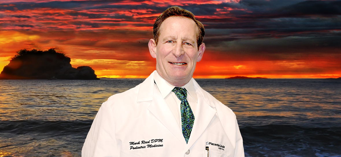
Mark Reed, DPM
DABFAS FAPWCA

714-528-3668


Podiatry Skin Diseases
Malignant Melanoma
Pigmented lesions should always be inspected and observed. Most pigmented areas are nothing but freckles and moles. However, a potentially deadly pigmented lesion that can occur on the foot and lower extremity is Malignant Melanoma.
These changes are usually subtle and may consist of increased size and depth of color, onset of bleeding, seepage of clear fluid, tumor formation, ulceration and formation of satellite pigmented lesions. The color is usually not uniform but is likely to be scattered irregularity, being brown, bluish black or black. An increase in pigmentation may precede enlargement of the lesion by several months. Although any part of the body may be affected, the most frequent site is the foot, then in order of frequency, the remainder of the lower extremity, head and neck, abdomen, arms and back. Malignant melanoma may also form under the nails of the feet and hands. The thumb and big toe are more commonly affected than the other nails. Quite often the adjacent skin to the nail is ulcerated. Usually, a fungal infection is suspected, and antifungal treatment may be administered for months before the true nature of the lesion is discovered. A black malignant melanoma of the toe can also be mistaken for gangrene. Overall, the incidence of malignant melanoma is quite low.
Actinic Keratosis
Another cancer-causing lesion that can occur on the feet are called Actinic Keratosis. Although most commonly found in sun-exposed areas of the body such as the face, ears, and back of the hands, these lesions can also occur on the foot. They are characterized as either flat or elevated with a scaly surface. They can either be reddish or skin colored. On the foot they are frequently mistaken for planter's warts. These lesions are the precursor of epidermoid carcinoma. Treatment for these lesions should be through as they are definitely precancerous. Treatment consists of freezing the lesions with liquid nitrogen or sharp excision.
Kaposi's Sarcoma
Yet another cancerous lesion that can occur on the foot is called Kaposi's Sarcoma. These lesions occur most commonly on the soles of the feet They are irregular in shape and have a purplish, reddish or bluish black appearance. They tend to spread and form large plaques or become nodular. The nodular lesions have a firm rubbery appearance. The appearance of these lesions is an ominous sign. In the late 1970's and early 1980's an outbreak of Kaposi's sarcoma occurred in San Francisco, California. It was later learned that the disease was associated with AIDS infection. It can occur without the concurrent AIDS infection, but this is very rare.
Chronic athlete's foot can cause an increased pigmentation to the bottom of the foot. It is associated with dry scaling skin and may have a reddish appearance.
Psoriasis
Psoriasis is a common, chronic, and recurrent inflammatory disease of the skin. It is characterized by round, reddish, dry scaling patches covered by grayish white or silvery white scales. The lesions have a predilection for the nails, scalp, elbows, shins and feet. On the feet, it can be difficult to distinguish it from athlete's foot, and the nail appearance may be confused with fungal infections of the toenails. The nail appearance does have a unique characteristic; it may have a pitting appearance. A characteristic feature of the condition is pinpoint bleeding when the scaled areas are brushed off. A variant of psoriasis is called pustular psoriasis. This form of the disease is characterized by small pustules or blisters filled with clear or cloudy fluid. This can mimic acute athlete's foot. It characteristically does not itch or burn. It is distinguished from athlete's foot by negative fungal cultures. The picture can become confusing because a secondary fungal infection is possible. In this instance both conditions are present at the same time.
Psoriasis can also affect the joints of the feet and lower extremities causing a painful arthritis. X-rays will show characteristic erosions of the bones in the toes. Treatment consists of anti-inflammatory medications, steroids, and other medications specific for the treatment of psoriasis.
Venous Stasis
Generalized increased pigmentation occurs for a variety of other reasons. Dark patches of skin occur about the ankles and lower legs in persons who suffer from Venous Stasis. Venous stasis is caused by an accumulation of fluid in the lower extremities. This is due to poor venous return of blood to the heart. Venous blood flow back to the heart occurs by way of the veins in the feet and legs. Venous stasis is associated with varicose veins that do a poor job of returning blood to the heart. As a result, the blood flow is slowed, becomes stagnant, and fluid accumulates in the ankles and lower legs. As the fluid accumulates in the lower legs, the small and medium-sized veins break or leak fluid into the tissues. As blood cells break up in the tissue, they deposit the iron that is part of hemoglobin in the blood cell. The iron stains the skin causing a light to dark brownish appearance. With time, the skin and subcutaneous fat becomes thinned and will break down creating weeping venous stasis ulcerations. At times, blistering will form with a clear, watery fluid weeping from the skin. This condition requires professional attention by a physician.
Diabetic Dermopathy
Another cause of generalized increased pigmentation is diabetes. The condition termed Diabetic Dermopathy occurs most frequently on the shins and lower legs. They may have the appearance of small scars. Their appearance may precede the diagnosis of diabetes by several years. The actual cause of diabetic dermopathy is not well understood, but it does not cause any particular problem or pose any particular health threat.
Small, spider-like areas of increased pigmentation on the ankles are caused by the breakdown of small veins in the area and are called Spider Veins; they also pose no health risks.
Blisters
Blisters form as a result of heat, moisture and friction. Blisters can also form as a result of fungal infections of the skin, allergic reactions or burns. If a patient has diabetes, they should be evaluated by a doctor in a timely fashion. Generally, a person will recognize a burn by association with a specific painful event. People with diabetes may not be able recognize the painful event due to a condition called neuropathy. A doctor should attend to burns. Blisters are due to fungal infection of the skin or to allergic reactions, which will generally occur in clusters and be smaller than blisters caused by friction. They will also often occur in areas of the foot, which are free from friction forces.
Blisters should be drained leaving the cover of the blister intact. The area should be protected with a non-stick bandage with mild compression. Ice to "hot spots" can be soothing and reduce the thermal damage to the surrounding area. "Double socking" can prevent blisters associated with athletics. Wearing two pair of socks allows the friction to be absorbed between the socks reducing friction to the skin. A sock has been developed that helps to reduce friction and blistering called the Thro-lo sock. It is useful for athletics and for diabetic patients. They are widely available in athletic shoe and apparel stores. Skin protectant sprays and adhesive gel pads are also available.
Abrasions
Abrasions to the skin are a result of excessive friction resulting in the partial loss of the epidermis. The area should be cleaned with an antibacterial soap and dressed in a non-stick bandage and a topical antibiotic ointment. It may take several weeks for the area to completely heal. During this period, the area should be protected from shearing forces. Deep abrasions can result in scaring. Any sign of infection should prompt a visit to the doctor.
Skin Tears
Skin Tears result from a rapid, forceful shear to the skin. Skin tears are most commonly self-inflicted by improperly removing adhesive dressings and tape. Careful counter pressure should be applied to the skin near the adhesive dressing as the dressing or tape is slowly removed. A common misconception is that paper tape will not damage the skin. To the contrary this tape can really stick to the skin and will tear the skin if removed improperly.
Mark Reed, DPM
DAPFAS FAPWCA
1275 N. Rose Drive, Suite 136
Placentia, CA 92870
Open M-F 8:30 to 5
Saturday: By Appointment
Info@Podiatry.Care
Fax: 714-528-0739
Office: 714-528-3668
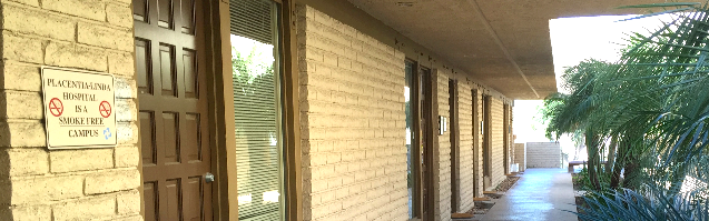
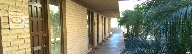
Located in the
Placentia Linda Hospital
Medical Building
Follow Dr. Mark Reed

*****
Beckie Burcham.
An absolutely amazing and genuine doctor who is not interested in only treating the symptoms, but rather treating the real cause! Dr. Reed explains everything in real simple terms that anyone can understand, especially seniors. He has amazing, common sense solutions (i.e. treatments) for unresolved diabetic foot ulcers where others have only wanted to continue cutting weekly into my family member's foot! Dr. Reed is absolutely AMAZING! And if you are getting no where with your currents foot issues or someone continues to only cut, please, please...do yourself a favor and contact Dr. Reed's office right away! Trust me, my family member has been dealing with an unresolved diabetic foot ulcer since 2018, and in just 3 weeks the foot is healing fantastic!!! Thank you Dr. Mark Reed. You have helped us to save the foot!!!
What an Amazing Podiatrist Dr. Mark Reed is! After years of other foot doctors just cutting into my family member's foot (diabetic foot ulcer), with numerous infections along the way...I did research and found Dr. Reed. Absolutely the best decision I have made for my loved one! After only 3 weeks of treatment, the foot ulcer is healing!!! No amputation! No continuous cutting and keeping the wound open and susceptible to infections! Brilliant man with great knowledge, kind heart, straight to the point and tells the truth the way it is! Dr. Reed does not just treat the symptoms, but rather the source of the problem! My family member's glucose levels are down to an average of 112 now. The wound is healing and new skin is growing!!! Amazing! I would highly recommend Dr. Reed to anyone and everyone! Thank you Dr. Reed, from the bottom of my heart! You have been a God send! Thank you!
*****
Mathew
H. I went to Dr. Reed because I had a chronic (5 months) wound on my leg that needed expert medical attention. Dr. Reed assessed the wound and knew how to treat it. He explained the process of healing the wound and what I needed to do in order to make the plan work.
I appreciated that Dr. Reed took the time to explain the process and his approach to me. As I followed the plan, I saw improvement within the first week and continued improvement in the following weeks. Within six weeks of my first appointment, the wound was healed.
Dr. Reed is personable and I enjoyed speaking with him in conversation. Most importantly, he told me what the treatment plan was, why it was that, and what I needed to do to ensure that it is effective. I am reluctant to go to doctors. I highly recommend Dr. Reed because he understands that many people feel that way and most importantly because the treatment was successful.
Lastly, the office staff are professional and kind. Dr. Reed's office is a first class group and I recommend them very highly.
*****
Paul Gordy. Dr. Reed is an outstanding podiatrist...he is state of the art medicine. This is by far the most kind, compassionate and caring physician who takes personal interest in his patients care and success in healing...when Care More and BlueShield doctors said they could not heal me...Dr. Read said "that's crazy", I'll heal you in 90 days .....and he DID!!!!!,
Amazing skill. You will not find a better person or doctor anywhere...
My family and I. Love Dr Reed.
You will too!
Paul Gordy
Yorba Linda Ca
*****
David M. The entire staff is friendly and very helpful. Dr. Reed is dedicated, spends time to take care of his patients. I had gangrene in my toe and Dr. Reed was diligent in his care and was able to eradicate the problem. I have recommended him to others, who had similar positive experiences.
***** Lee Godden. I have nasty bunions which eventually led to a painful stress fracture of the second metatarsal while playing basketball 2x/week. Dr. Reed managed my entire recovery, from initial visit to discharge two months later. He took X-rays and did ultrasound therapy. I was happy that his focus was on realignment and patient education, as opposed to surgery. I'm now running again thanks to the orthotic insoles, toe spacers, etc. that Dr. Reed put me in. I highly recommend Dr. Mark Reed.
***** Yvon Cariou. Dr. Reed did a great job on that ingrown toe nail, a painful condition. It took some time but Dr. was persistent and supported the process with detailed explanations and supplemental care as needed. A good office to go to.
***** Sue Alexander. If you’re looking for a podiatrist (foot and ankle doctor), may I suggest Dr. Mark Reed in Yorba Linda. Throughout my treatment Dr. Reed explained what he would be doing and why. Thank you Dr Reed for your expertise, kindness and encouragement! Sincerely, Sue Alexander
***** Salam Hamad. Hat off for Dr. Reed. He was the 4th doctor, second specialist to consult for my Plantar Fasciitis case. I went to him after the 1st specialist, using injections, boots and what not decided to operate on my heel in 30 days if the pain does not go away. Doctor Reed threw all that talk away and dismissed every recommendation. I went to him armed with lot of documents, dates and details of what the first 3 doctors did. He said, don't give the history, just tell me where it hurts. He examined the heel and the foot and diagnosed the problem immediately. He then prescribed the method to treat it. He did it! yes, it took more time than he thought but he DID it. I owe doctor Reed and appreciate him, his knowledge, experience and his caring attitude every time I walk. Yes, walking became possible for what he did. Oh! by the way, he is not in it for the money.
***** Sue Alexander. If you’re looking for a podiatrist (foot and ankle doctor), may I suggest Dr. Mark Reed in Yorba Linda. Throughout my treatment Dr. Reed explained what he would be doing and why. Thank you Dr. Reed for your expertise, kindness and encouragement! Sincerely, Sue Alexander
***** Monsie Crane.
Dr Reed cured me of the neuropathy in my toes. The neuropathy was a result of chemotherapy, which I received as a cancer treatment. He also helped me with proper shoe fitting for bunions and longer second toes. I highly recommend Dr Reed.
***** Colleen Livermore. Dr Mark Reed has been an excellent doctor, evaluating my foot and ankle pain, and giving me the instruction and exercises needed to strengthen and heal. I appreciate his excellence, expertise and experience. Ankle pain can be so challenging so to feel better after his help is a great relief.
Reply1
*****
Matthew Jones.
Dr. Reed has been my podiatrist for approximately 2 years now. He remains one of my favorite doctors because of how kind he is, how punctual he is and most importantly how effective he is.
I have been to several other podiatrists in the past and I just about gave up until I found Dr. Reed on google. The results he has given me have changed the way I feel and the way I look.
***** Lily Lambutu. Dr. Reed is an excellent doctor. He listens, gets straight to the point, explains everything, and pays attention to detail. He answers all questions honestly. I've seen him for ingrown toenails and a tumor on the bottom of my foot. I trust him completely as he takes the time to assess the issue and will take a conservative approach if it will solve the problem.
***** Bryant Brislin. Dr. Reed took a thoughtful approach to my issue and came up with a solution that other podiatrists had not. He took his time and was very friendly, which was refreshing. He seemed to truly care for me to get better!
***** Lisa Fogerty.
Dr Reed is an outstanding doc. Several years ago I had a hammer toe surgery done by another doctor who damaged the nerves in my foot. I was in pain for 3 years and was told by that doctor that I would have to live with it and that eventually the nerves would heal. They didn't. I couldn't sleep due to the pain and had to alter my exercise routine. Dr Reed took on the case, correctly diagnosed the problem and started a program of injections to get the nerves to regenerate. He told me to be patient and that "we" would fix the problem. He always kept me informed of the procedures and made me part of treatment plan. It took several months but today my foot and toes are healed thanks to Dr Reed's persistence.
My wife dislocated several toes in an accident and was in significant pain. Dr Reed fixed her up as well.
We are both very grateful to Dr Reed and his excellent staff for their kind and professional medical services and highly recommend him. If you want the best podiatrist in Orange County you found him in Dr Mark Reed.
***** Samantha Smith. It’s been almost 2 years since Dr. Reed cut off an amputation stump neuroma and then reconnected my husbands medial plantar nerve. This man has changed our lives forever. After being turned down and rejected by SO many doctors, we had no hope left...then we found Dr. Reed. He really did the impossible! My husband was in so much pain for so long and now feels little to no pain thanks to our friend Dr. Reed. We owe everything to him and will always be SO thankful for everything he has done for us. He will forever be a huge part of our lives.
Reply1
***** Leo Burke. I'm an avid hiker but have been plagued for years by a large toenail that was sensitive to pressure and would have to go to great lengths to avoid losing the nail. But every now and then I would lose the nail and all this was impacting my hiking (and ruining some vacations). I was seeing other podiatrists and we would try various work-arounds, but I finally decided that I would get rid of that nail once and for all. I had a lot of trepidation about that, but I decided to see Dr. Reed to consult with him on doing so. He assured me that it would be no problem at all, which surprised me, so I agreed to have him do the removal. Amazingly, it turned out to be a piece of cake with little to no pain, and I was back in action very quickly. It pains me to realize that if I had seen him a long time ago I could have avoided years of aggravation and frustration. Not only that, but he's an expert on hiking boots and given me a lot of help in boot selection.
He's been in this for so long that he's a fountain of knowledge and enjoys imparting his wisdom to his patients. For example, I also have a pressure sensitive small toe on my other foot and he was able to give me samples of some products that can relieve pressure on the toe that I hadn't been aware of. If that doesn't do it, I can cut an "X" in the boot over that area and cover it with a piece of leather. He then supplied me with a piece of leather that could be used for that purpose and told me what glue to use.
There's no question about it - Dr. Reed is the man to see for your foot problems. He's head and shoulders above everyone else.
***** Gloria Gonzalez. Dr. Reed is a great podiatrist. He's the only doctor that has helped my family's and my foot issues. He didn't suggest surgery for my issue, and his solution was easy and innovative. A couple of years later, and his solution still works.
Reply1
***** Nikki Clark. I have been a patient of Dr. Reed's for 6 months. During this time I have found both his professional staff and Dr. Reed to apply the utmost patience, compassion and expertise. They always make time for appointments and during doctor visits. Dr. Reed has an impeccable bedside manner, provides all options available and genuinely cares for a positive solution. Dr. Reed is a student of his practice and applies current medical advancements. I would recommend Dr. Reed and his staff to family and friends for problems they might be experiencing.
*****
Viet Tieu Bao: I have a great experience with Dr. Reed. First of all, he's an awesome surgeon as it turned out i didn't have any pain after my surgery (i had a tumor that grew next to my Achilles tendon that needed to be removed and in order to have a clean removal of the tumor so it won't come back Dr. Reed had to shave off 1/3 of my Achilles tendon). Amazingly i did not have to take a single pain killer. Hats off to his skills. Secondly, his caring is extremely important since he encouraged me to call him anytime if i have a concern or just a question. I did call him on the day of the surgery i noted the time 11:30pm and he did return my call just few minutes later. I was amazed how pleasant and calm he was even though that late in the night and he ensured me that i feel ok before he let me go back to sleep. Last but not least, he is very detail oriented surgeon as he always explained clearly his procedure & options & pros & cons as well as any medical terms that i don't understand. I feel i was so lucky that I chose Dr. Reed for my problem and i highly recommend anyone who needs a DPM should give it a try with Dr. Reed. Am confident that you won't be disappointed.
Thanks Dr. Reed for taking good care of me and deeply appreciated for all you did for me.
***** Uli H. Went to Dr. Reed about 2 months ago because I woke up with excruciating pain on top of my foot. There was no injury or even exercise the day before so it was totally random. I explained this to Dr. Reed and he diagnosed it as tendinitis. He explained to me why it happens and it made perfect sense. So in one session I basically found out that I've been wearing the wrong shoes for my flat feet this whole time. What a difference my life would've been if I had visited him years ago. He told me my knees are not aligned correctly because of my flat foot which explains my knee pain. He referred me to a place to get some better walking shoes and showed me creases on my shoes that showed just how much pressure is on one side of my foot. The custom orthotics he made me have been a life changer. The only way I can describe the feeling is I use to feel like I was walking on unstable stilts and now my feet have custom made platforms and I'm stepping on a solid foundation every step. The night splint he gave me was the solution to the tendinitis. It is now gone and even played soccer within a month of first going. I'm now working on my pre-existing ankle injury and have to test out my tendonitis ankle to make sure its completely gone. Dr. Reed is great, has some great advice and I definitely recommend him.
***** Douglas Ho. I am a returning customer after many years. Returned to Dr Reed to evaluate my Plantar Fasciitis. He recommended stabilizing tennis shoes and a night boot to keep my foot in position. He said I have a mild bursitis on my heel. His recommendations are spot on and I am slowly recovering. His background in Sports medicine was also helpful in commenting on a treatment for my tennis elbow. I love it.
***** Richard Baugh. Dr Reed has helped me excellently over the years to overcome several different foot problems: ingrown toenails (his treatment was painless!), neuroma and plantar fasciitis. He is friendly and takes the time to explain causes and treatments. I’m most grateful to him and will continue to use his services.
**** Victor Skillings. It was a phenomenal experience from the initial consultation, pre-surgery preparation, surgery and follow-up visits. Now, I know why so many of his patients feel compelled to write great reviews about Dr. Reed. His approach to patient care, willingness to explain the procedure and answer all your questions, surgical skills and genuine interest in his patients were far, far better than what I had expected.
I had bunion surgery about two months ago. It was extremely large, about the size of a walnut. My procedure took a little more than an hour, and to my amazement, I felt absolutely no pain during or anytime after the surgery. In a few weeks, I was able to walk fairly normally.
In summary my review is five out of five stars, thumbs up, A+, highly recommended and all the above! So if you’re looking for the best foot surgeon in Orange County, look no further – Dr. Mark Reed should be your first and only call.
***** Juan Vallejo. Okay I'm a very critical. This Dr and his office staff are hands down the best. I had a great experience with Dr. Reed. I have recommended him to all the people I know. Everyone is great and professional.
***** Karina Lerma. He is the BEST Podiatrist in Orange County. He truly cares for the patients and tries to give them the best treatment!
© 2015 PLFAG. All rights reserved.

***** Juan V in Canyon Country, CA. Okay I'm a very critical. This Dr and his office staff are hands down the best. I had a great experience with Dr. Reed. I have recommended him to all the people I know. Everyone is great and professional.
***** Laila in Yorba Linda, CA. After visiting 2 other doctors in a period of 2 years, with no success. Dr. Reed was able to pinpoint the problem with my feet! Finally, I have no pain while running and working out.
***** Pam L in Villa Park, CA. I am so pleased with the results of Dr. Reed's care of my daughter's foot pain while wearing her figure skates. She has figured skated for a few years, and recently developed arch pain while wearing her skates that would not go away no matter what she did. He made custom orthotics to fit in her skates and support her feet, and she is currently pain free! If you have an athlete or figure skater, I would definitely recommend Dr. Reed for podiatry care.
***** J. Barna in Blue Jay, CA. I've had an amazing experience with Dr. Reed and his office staff! I was in significant pain from a neuroma when I first came to his office. Dr. Reed was my second opinion, because the first office, turns out, had no idea what they were doing. Dr. Reed was able to reduce my pain until I needed surgery. After a short recovery I am now near 100%!! I would recommend Dr. Reed to anyone in Southern California that is suffering from any type of foot appointment.

***** Cher Corso. My fourteen year old daughter (a dancer) was having problems with both feet. We had already been to one podiatrist with no satisfactory resolution when we found Dr. Reed. The entire experience was positive for our entire family. Dr. Reed knew exactly what was going on and corrected the problem. I have already recommended Dr. Reed to a very dear friend who's son is having foot problems. This is the place to go! Great experience for my daughter who was scared of doctors before seeing Dr. Reed.
**** May Liping. I had a painful bump on the top of my foot for months. My previous podiatrist, misdiagnosed me and scared me half to death. First he claimed my ganglion cyst was caused by my wearing bad shoes. Then, when my foot continued to hurt and I saw him again, he claimed the cyst "had hardened" and therefore, only surgery could remove it. He made this declaration without even x-raying my foot. Scared, I went to Dr. Reed for a second opinion and thank God I did. Dr. Reed did an x-ray and determined that the cyst could be treated immediately. He looked stunned when I told him about the other doctor's diagnosis. He told me that, soft or hard, cysts can be taken care of. He gave me an injection of steroids and the cyst has disappeared ever since. Dr. Reed saved me from unnecessary surgery and for that, I will always be grateful!
***** Linda Rodgers. Dr. Mark Reed was the second doctor I went to for my ingrown toenail. The first foot doctor did not do the procedure right and the ingrown nail returned. The pain from the procedure was also terrible and I do not think the doctor understood what she was doing because she was saying that it is supposed to hurt. Dr. Reed took a lot of time to explain the procedure and reassure me that the injection was going to go smooth. Dr. Reed used a skin vibrator that made the shot painless and I did not feel anything as to the needle and the numbing of my toe. Dr. Reed talked about a number of interesting topics so the procedure was done and felt like it only took a few minutes. The office is very clean and the staff is great. There is not a better doctor out there for foot care. I highly recommend that you go to this doctor.